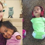
TWO-YEAR-OLD GIRL WITH HYDROCEPHALUS UNDERGOES SUCCESSFUL SURGERY TO REDUCE HER ENLARGED HEAD IN A LIFE
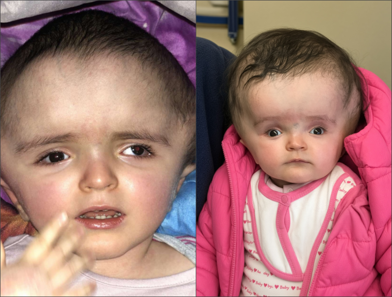
A remarkable medical breakthrough gave a 2-year-old girl a new chance at life after surgeons reconstructed her skull, which had become extraordinarily enlarged due to hydrocephalus.
A little girl born into a family in North Africa had a head that was unusually large in proportion to her body. During pregnancy, ultrasound scans revealed her condition, allowing her family to understand the challenges she might face.
Hydrocephalus caused her skull to enlarge abnormally as fluid gradually accumulated inside her brain.
As the pregnancy progressed, doctors warned that if her parents chose to continue, she could face difficulties with feeding and mobility, severe developmental challenges, or potentially life-threatening complications shortly after birth.
Because her skull was unusually enlarged, she could not be delivered vaginally. Doctors recommended a Caesarean section at just 34 weeks of pregnancy. She was born safely, but the medical resources available in Africa did not allow for further intervention to help her.
By May, her parents decided to take their daughter to Sainte-Justine Hospital in search of treatment when she was nine months old. Upon arrival, doctors immediately recommended that she be admitted to the emergency department.
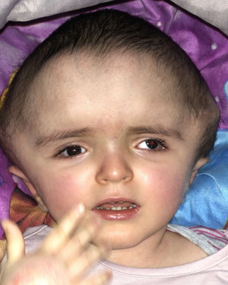
According to Dr. Alexander Weil, the little girl had an enormously enlarged head filled with fluid, and her scalp had become so stretched and thin that the veins were clearly visible. Her skull bones were also beginning to separate. With her head measuring 76cm in circumference — nearly twice the size of an average child’s head at her age — she could move her arms and legs but was unable to roll over, crawl or sit up.
Hydrocephalus is a neurological condition that can become life-threatening, particularly in countries with limited medical resources. In developed countries, early intervention can often help manage the condition and prevent severe complications.
The condition occurs when cerebrospinal fluid cannot circulate properly through the four spaces within the brain. As pressure builds, the skull expands like an inflating balloon. A child’s head can grow rapidly during the first years of life, when the brain and skull are developing at their fastest rate.
The first treatment involved targeting the structures responsible for producing cerebrospinal fluid, but it was unsuccessful. Doctors then inserted a shunt into the base of the girl’s skull to drain the fluid. Although the fluid was brought under control, the size of her head remained extraordinarily large.

A remarkable breakthrough after 12 hours of painstaking surgery
Dr. Alexander Weil, Daniel Borsuk and Dominic Venne, the surgeons responsible for her care, proposed a bold procedure to reshape and reduce the size of her skull by cutting and repositioning the bones. It was considered the most promising option available, but it also carried significant risks, including severe bleeding, stroke and infection. Her parents agreed to give their daughter a chance.
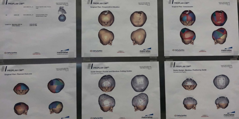
Using CT scans of the girl’s skull, doctors created a 3D model. They mapped out the cuts and reconstructed the skull on the model to simulate how it would look after surgery, allowing them to carefully plan the procedure.
The operation began when the girl was 26 months old. Surgeons made an incision across her scalp to expose the skull, then carefully cut the bones into multiple sections according to the measurements established on the 3D model.
At the same time, they drained nearly three litres of fluid from her brain — compared with approximately 150ml in a typical person. The amount was carefully calculated to reduce pressure without damaging veins or triggering excessive bleeding.
As the fluid was removed, the brain began to shrink. Surgeons meticulously removed excess tissue, trimmed and repositioned the skull fragments, reconstructed the smaller skull and secured the bones with screws. After 12 hours of incredibly precise work, the operation was successfully completed.
Following surgery, the little girl remained unconscious for 24 hours. One month later, her head was significantly smaller, although it was still larger than average. Her surgical scars had healed and were barely visible. She was able to play, smile and laugh with her family again. Doctors explained that it could take years for her head to gradually approach a more typical size.
Like other children living with hydrocephalus, she will need rehabilitation and physical therapy to strengthen her neck muscles and improve her mobility. Other potential complications remain uncertain, and she will require ongoing medical monitoring.