SEEING THE INVISIBLE: THE EVOLUTION OF FLUORESCENCE-GUIDED SURGERY
SEEING THE INVISIBLE: THE EVOLUTION OF FLUORESCENCE-GUIDED SURGERY
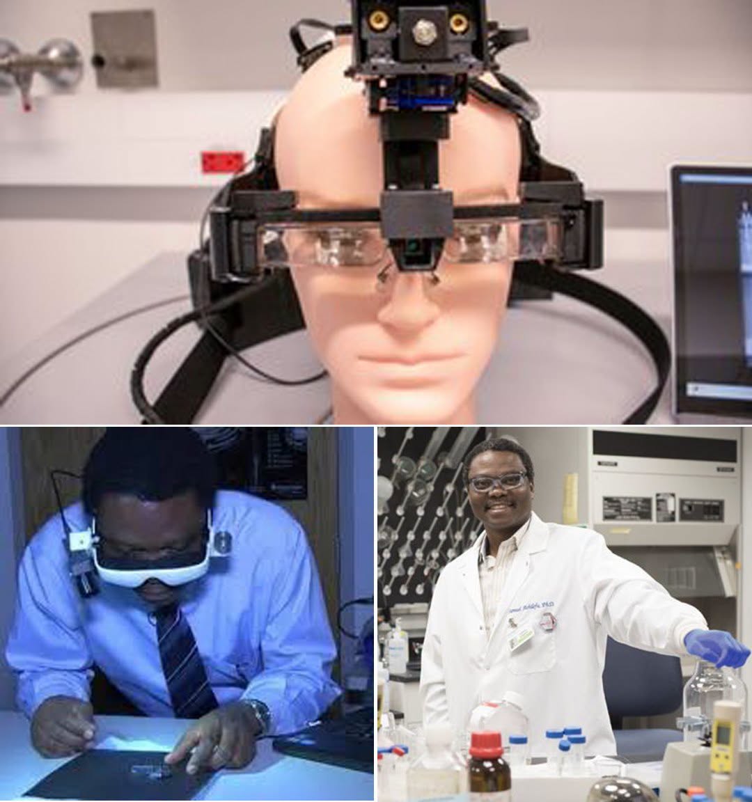
In 2014, a surgeon put on a pair of specialized goggles and saw something the human eye normally cannot detect: fluorescent signals emanating directly from tissue inside a patient.
This innovative technology was developed by a team led by scientist Samuel Achilefu at Washington University in St. Louis. The underlying principle was simple yet powerful: combine a fluorescent agent with near-infrared light to help medical teams visualize critical tissue that would otherwise be difficult to distinguish during an operation. Because near-infrared light is naturally invisible to human vision, the custom goggles were designed to capture that signal and instantly convert it into a visible image directly within the surgeon’s field of view.
On February 10, 2014, the technology made its clinical debut at Siteman Cancer Center. Breast surgeon Julie Margenthaler wore the system while operating on a breast cancer patient. The goal was not to replace the surgeon’s judgment or magically detect every single cell, but to provide real-time visual assistance during surgery, helping clinicians better identify marked areas that required precise removal.
Researchers continued refining the concept over subsequent years. By 2024, a new system named FAR-Pi took the idea a step further. Built using low-cost Raspberry Pi microcomputers, standard optical parts, 3D-printed components, and augmented-reality glasses, FAR-Pi demonstrated a cheaper, more portable approach to fluorescence-guided imaging in preclinical trials.
Advanced medical equipment is often prohibitively expensive and difficult to deploy in under-resourced hospitals. If portable and affordable imaging tools can reach widespread clinical reliability, surgeons everywhere could soon gain a clearer view of hard-to-see tissue, ensuring that technology invisible to the eye helps guide life-saving decisions in the operating room.