SEPARATED SURGICAL REALITY: HOW 3D TECHNOLOGY SAVED TWO CONGENITAL TWINS

SEPARATED SURGICAL REALITY: HOW 3D TECHNOLOGY SAVED TWO CONGENITAL TWINS
Before surgeons ever separated Minal and Mirha, they had already practiced the complex operation on a digital counterpart. The twin girls were born in Pakistan with a rare condition called craniopagus, meaning they were joined at the head. Their connection extended beyond bone, sharing critical blood vessels and brain tissue. Any single mistake could severely damage the blood supply to either child’s brain.
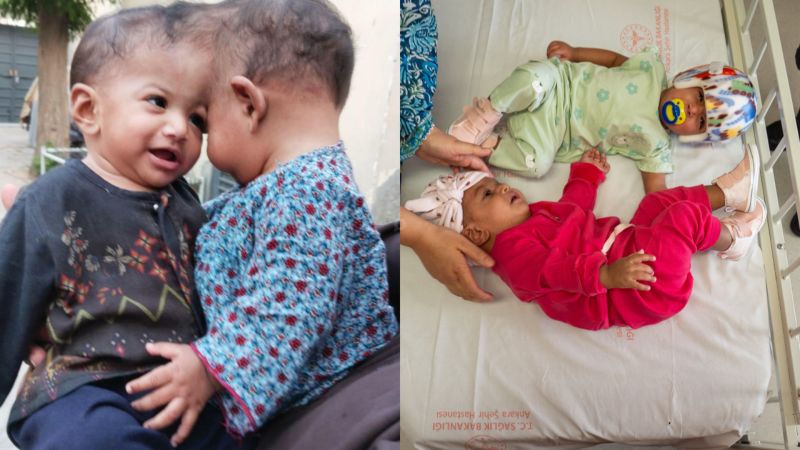
To mitigate these risks, British neurosurgeon Professor Noor ul Owase Jeelani and an international medical team utilized high-definition digital modeling. Using advanced imaging scans, specialists built a precise 3D model of their anatomy. Mixed reality technology enabled surgeons across the globe to examine shared structures and rehearse the procedure virtually prior to making a physical incision.
The ultimate separation took place across two surgical stages at Ankara Bilkent City Hospital in Turkey. On July 19, 2024, a team of roughly 60 medical professionals initiated the final 14-hour operation. Surgeons successfully divided the shared structures, granting both girls distinct physical independence. Two months following the procedure, the medical team confirmed that both children were recovering well and preparing to return home to Pakistan.

This innovative approach builds upon previous successes, including the 2022 separation of Brazilian twins Bernardo and Arthur Lima. By integrating mixed reality, specialists from different continents collaborated on complex anatomical maps in real time, setting a new standard for high-risk pediatric surgery.