UNDERSTANDING CROUZON SYNDROME: SURGERY, DISTRACTION, AND RECONSTRUCTION
UNDERSTANDING CROUZON SYNDROME: SURGERY, DISTRACTION, AND RECONSTRUCTION
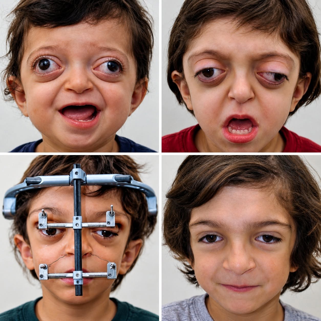
Crouzon syndrome is a genetic craniosynostosis disorder, usually tied to mutations in the FGFR2 gene, where skull bones fuse prematurely. This early fusion restricts normal skull and facial growth, causing midface hypoplasia—a severe underdevelopment of the middle face. Consequently, shallow eye sockets leave eyes prominent and unprotected, while a receded upper jaw causes severe airway obstruction, sleep apnea, and bite misalignments.

Addressing severe cases requires a multidisciplinary craniofacial team. Beyond traditional surgery, a key technique for midface advancement is distraction osteogenesis. Doctors perform a Le Fort III osteotomy to separate the underdeveloped midface bones, then attach a specialized external metal frame. Rather than moving bones all at once, the device gradually separates bone segments by roughly 1 millimeter per day. As the space widens, the body naturally generates new bone in the gap. Clinical cases show midface advancements ranging from 10 to 27 millimeters, drastically expanding the airway, improving jaw position, and protecting vision.
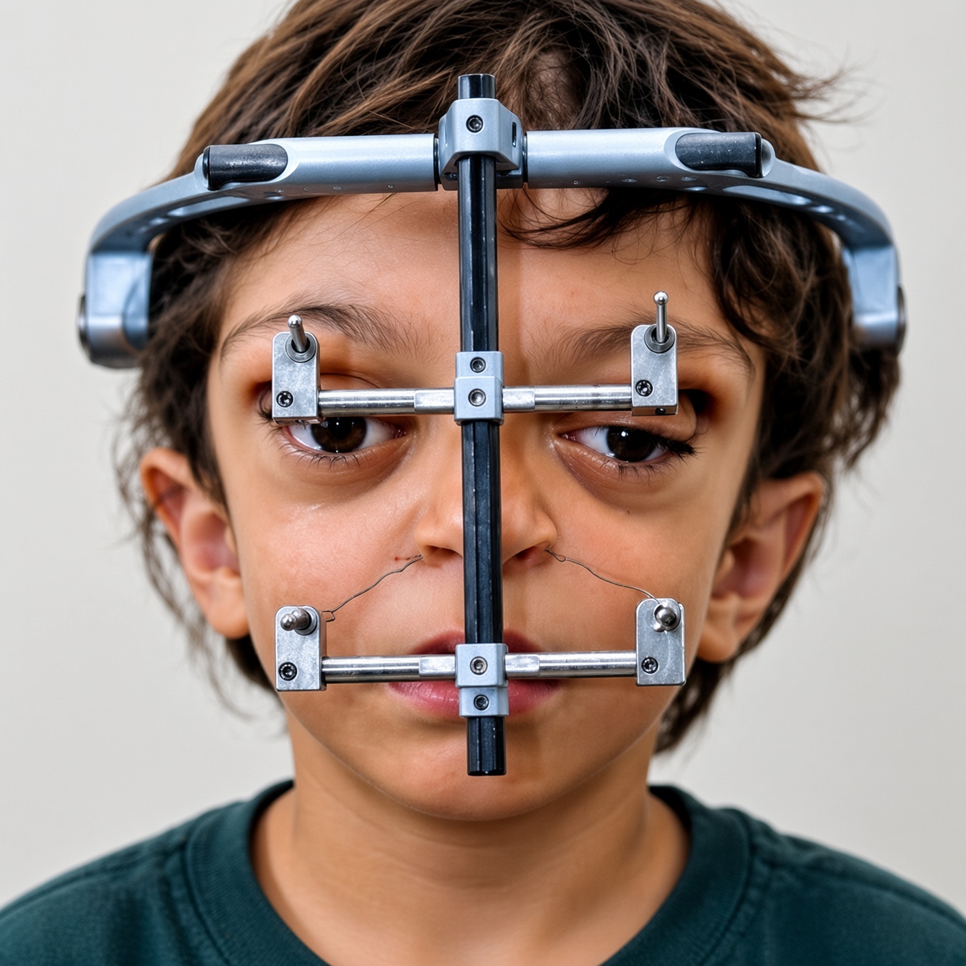
Following the advancement phase, patients enter a consolidation period of around three months, allowing the newly formed bone to solidify before removing the device. Long-term monitoring remains essential as the child continues to grow. The metal frame is not a new face, but a precise tool enabling the body to rebuild itself—restoring vital function and structural support.